
Report findings at diagnosis of the primary disease for infusion. All values reported must reflect testing performed prior to the start of first treatment of the primary disease for which the HCT / cellular therapy is being performed. If multiple studies were performed prior to the start of therapy, report the assessment closest to the diagnosis date of the primary disease for infusion. If the recipient’s MDS transformed, report the studies from the original diagnosis.
Question 18: Date CBC with differential tested
Report the date the sample was collected for testing and continue with question 19. If multiple studies were performed prior to the start of therapy, report the assessment closest to the diagnosis date of the primary disease for infusion.
Questions 19-20: Neutrophils
Indicate whether the neutrophil percentage in the blood was “known” or “unknown” at the time of MDS diagnosis. If “known,” report the value documented on the laboratory report in question 20. If “unknown,” continue with question 21.
Questions 21-22: Bands
Indicate whether the band percentage in the blood was “known” or “unknown” at the time of MDS diagnosis. If “known,” report the percentage documented on the laboratory report in question 22. If “unknown,” continue with question 23.
Questions 23-24: Metamyelocytes
Indicate whether the percentage of metamyelocytes in the blood was “known” or “unknown” at the time of MDS diagnosis. If “known,” report the percentage documented on the laboratory report in question 24. If “unknown,” continue with question 25.
Questions 25-26: Myelocytes
Indicate whether the myelocyte percentage in the blood was “known” or “unknown” at the time of MDS diagnosis. If “known,” report the percentage documented on the laboratory report in question 26. If “unknown,” continue with question 27.
Questions 27-28: Lymphocytes
Indicate whether the lymphocyte percentage in the blood was “known” or “unknown” at the time of MDS diagnosis. If “known,” report the percentage documented on the laboratory report in question 28. If “unknown,” continue with question 29.
Questions 29-30: Monocytes
Indicate whether the monocyte percentage in the blood was “known” or “unknown” at the time of MDS diagnosis. If “known,” report the percentage documented on the laboratory report in question 30. If “unknown,” continue with question 31.
Questions 31-32: Basophils
Indicate whether the basophils percentage in the blood was “known” or “unknown” at the time of MDS diagnosis. If “known,” report the percentage documented on the laboratory report in question 32. If “unknown,” continue with question 33.
Questions 33-34: Eosinophil
Indicate whether the eosinophil percentage in the blood was “known” or “unknown” at the time of MDS diagnosis. If “known,” report the percentage documented on the laboratory report in question 34. If “unknown,” continue with question 35.
Questions 35-36: Was a bone marrow examination performed?
Indicate if a bone marrow examination was performed at the time of MDS diagnosis. If “yes,” indicate the date the sample was collected in question 36. If “no” or “unknown,” continue with question 40.
Question 37: Cellularity
Cellularity describes the percentage of bone marrow occupied by hematopoietic cells compared to other tissues, such as adipose (fat) cells. In MDS, the percentage of hematopoietic cells is likely increased (hypercellular) due to proliferation of immature cells. In other cases, the cellularity may be normal (normocellular) or decreased (hypocellular). This distinction is made on the pathology report of a bone marrow examination.
Indicate whether the bone marrow examination revealed “decreased (hypocellular),” “normal (normocellular),” or “increased (hypercellular)” cellularity at diagnosis or prior to the first treatment. If the degree of cellularity is not addressed in the report, select “unknown.”
Question 38: Myelofibrosis grading by WHO classification
Fibrosis describes the replacement of bone marrow by fibrous (scar) tissue. This distinction is made on the pathology report of a bone marrow examination and the myelofibrosis grade may be documented by the pathologist.
Indicate if the myelofibrosis grading is “known” or “unknown.” If the myelofibrosis grade is documented on the pathology report, select “known,” continue with question 39. If the pathology report is not available and the grade is documented in a physician note, then this would be sufficient.
If the myelofibrosis grade is not documented in the pathology report, select “unknown,” continue with question 40.
Question 39: Specify the grade
Specify the Myelofibrosis grading using the WHO classification. The classification and results should be clarified in the pathology report as dictated by the pathologist.
Select “MF-0” if the report documents scattered linear reticulin with no intersection (crossovers) corresponding to normal BM.
Select “MF-1” if the report documents a loose network of reticulin with many intersections, especially in perivascular areas.
Select “MF-2” if the report documents diffuse and dense increase in reticulin with extensive intersections, occasionally with local bundles of thick fibers mostly consistent with collagen, and/or focal osteosclerosis.
Select “MF-3” if the report documents diffuse and dense increase in reticulin with extensive intersections and coarse bundles of thick fibers consistent with collagen, usually associated with osteosclerosis.
Question 40: Were molecular tests for molecular markers performed (e.g., PCR)
Molecular assessment involves testing blood or bone marrow for the presence of known molecular markers associated with the recipient’s disease. Molecular assessments are the most sensitive test for genetic abnormalities and involve amplifying regions of cellular DNA by polymerase chain reaction (PCR), typically using RNA to generate complementary DNA through reverse transcription (RT-PCR). The amplified DNA fragments are compared to a control, providing a method of quantifying log increase of genetic mutation transcripts. Each log increase is a 10-fold increase of gene transcript compared to control. Molecular methods are evolving and now include chromosomal microarray / chromosomal genomic array, Sanger sequencing, and next generation sequencing (e.g., Illumina, Roche 454, Proton / PGM, SOLiD).
Indicate if molecular studies were obtained at the time the recipient was diagnosed with MDS or prior to the start of treatment.
If molecular studies were performed, select “yes” and continue with question 41.
If no molecular studies were obtained or it is unknown if molecular studies were performed, indicate “no” or “unknown” and continue with question 48.
Question 41: Indicate if a positive molecular marker(s) was identified
Indicate if a positive molecular marker was identified at the time of MDS diagnosis or prior to the start of treatment.
If a positive molecular marker was identified, select “yes” and continue with question 42.
If there were no molecular markers identified, select “no” and continue with question 47.
Question 42: Date sample collected
Report the date the sample was collected for molecular testing. If multiple studies were performed prior to the start of therapy, report the last assessment prior to the start of treatment.
Questions 43-44: Specify the positive molecular marker
Specify the positive molecular marker and continue with question 45. If a positive marker is detected, but not listed as an option, select “Other molecular marker” and specify the positive molecular marker in question 44.
Questions 45-46: Amino acid change
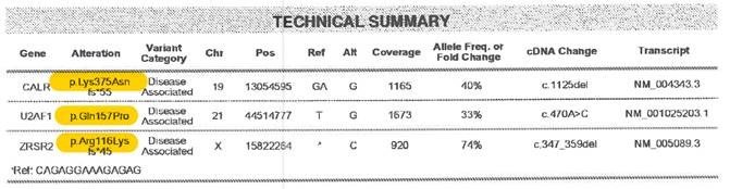
Amino acid changes can be described using standard mutation nomenclature. For CIBMTR reporting purposes, only amino acid changes based on protein-level sequences, beginning with the prefix “p.,” need to be reported. See Figure 1 below for an example of an amino acid change within a molecular pathology report.
Figure 1. Molecular disease assessment with amino acid changes documented (highlighted in yellow).
For more information on nomenclature used for amino acid changes, visit the Human Genome Variation Society Sequence Variant Nomenclature Protein Recommendations.
For question 45 indicate if the amino acid change is “known” or “unknown” for the positive molecular marker reported in questions 43-44. If known, report the amino acid change in question 46. If unknown, continue with question 47.
Copy questions 43-46 to report more than one positive molecular marker.
Question 47: Was documentation submitted to the CIBMTR?
Indicate if the molecular report(s) is attached to support the molecular findings reported in questions 43-46. For further instructions on how to attach documents in FormsNet3SM, refer to the Training Guide .
Section Updates:
| Question Number | Date of Change | Add/Remove/Modify | Description | Reasoning (If applicable) |
|---|---|---|---|---|
| 18 | 4/21/2023 | Modify | Clarified the instructions for the labs at diagnosis should reflect the labs closest to the date of diagnosis. | To ensure more consistent reporting, all labs for the “at diagnosis” timepoint were clarified to reflect the values obtained closest to the date of diagnosis, prior to the start of any therapy. |
Need more help with this?
Don’t hesitate to contact us here.